Cardiac Computed Tomography for Atrial Volume Estimation
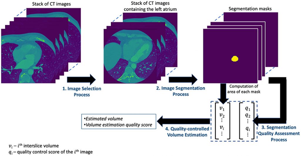
Using a dataset of 85,477 Cardiac computed tomography (CCT) images from 337 patients, a framework consisting of several processes that perform a combination of tasks is proposed. These tasks include the selection of images with full volumetric left atrium (LA) from all other images using ResNet50 (a residual deep learning neural network model with 50 layers), the segmentation of images with LA using UNet (a convolutional neural network image segmentation model), the assessment of the quality of the image segmentation task, the estimation of a larger LA volume (LAV), and the quality control (QC) assessment.
The proposed LAV estimation framework achieved accuracies of 98% in the image classification task, 88.5% in the image segmentation task, 82% in the segmentation quality prediction task, and R2 (the coefficient of determination) value of 0.968 in the volume estimation task. It correctly identified 9 out of 10 poor LAV estimations from a total of 337 patients as poor-quality estimates.
To read more: https://doi.org/10.3389/fcvm.2022.822269

